
Common Eye Disease
Age Related Macula Degeneration (AMD)
Understanding changes that can affect your central vision
What is Age Related Macula Degeneration?
Age-related macular degeneration, usually shortened to AMD, is a condition that affects the macula, the small, highly specialised area at the centre of the retina responsible for our clearest, most detailed vision.
The macula is what allows you to read, recognise faces, see fine detail and appreciate colour. AMD causes changes within and beneath this delicate area of the retina. It becomes more common as we get older and can affect one or both eyes, sometimes differently.
There are two main forms of AMD: dry AMD, which usually develops gradually, and wet AMD, which can cause more rapid changes in vision and requires prompt assessment and treatment.
AMD affects central vision rather than peripheral vision, so it does not usually cause complete blindness. However, if it progresses, it can make everyday activities that depend on detailed vision increasingly difficult.
Importantly, early AMD may cause no noticeable change in your sight at all. Changes can sometimes be seen during an eye examination before you are aware of any symptoms.
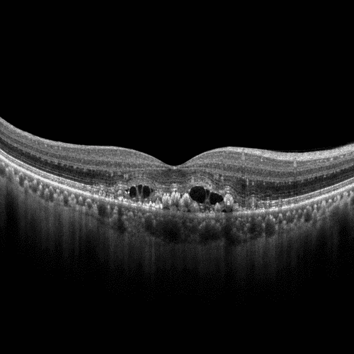
What you might notice
AMD can affect people in different ways, and the symptoms depend on the type and stage of the condition. In its early stages, you may notice no change in your vision at all. As AMD progresses, changes tend to affect the detailed central vision you use for tasks such as reading and recognising faces.
Some of the changes you might notice include:
What is happening inside the eye?
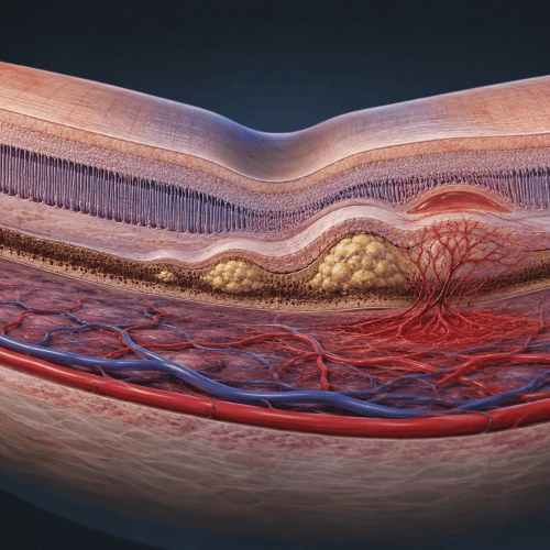
In AMD, changes develop in and around the macula, the part of the retina responsible for your detailed central vision. One of the earliest signs can be the appearance of small deposits called drusen, which collect beneath the retina. Small drusen are common as we get older, but larger or more numerous drusen can be associated with AMD.
In dry AMD, there can also be gradual changes to the cells that support and nourish the retina. Over time, these changes may affect the light-sensitive cells of the macula and, in more advanced disease, areas of these cells can become damaged or lost.
In wet AMD, abnormal new blood vessels develop beneath the retina. These vessels are fragile and can leak fluid or blood, disturbing the delicate structure of the macula and sometimes causing a sudden change in central vision.
This is one reason OCT imaging is so useful. It allows us to look through the individual layers of the macula and see changes beneath the surface that may not be obvious from looking at the retina alone.
SEEING BENEATH THE SURFACE
What is happening inside the eye?
OCT imaging
Some of the changes associated with AMD happen within or beneath the layers of the macula, where they cannot always be fully appreciated simply by looking at the surface of the retina.
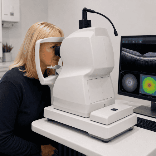
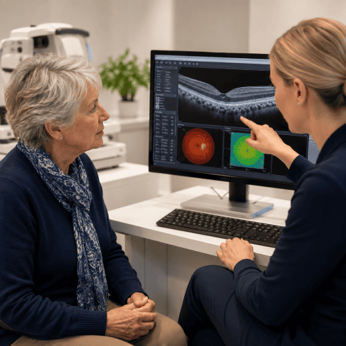
OCT imaging gives us a highly detailed cross-sectional view through these layers. It can show changes such as drusen, disruption of the retinal layers and the presence of abnormal fluid, sometimes before there is a noticeable change in vision.
Just as importantly, OCT gives us a detailed record that we can compare with future scans. This allows us to look for subtle changes over time and identify when something that has previously been stable may need closer investigation.
The bigger picture with Optomap
A wider view with Optomap
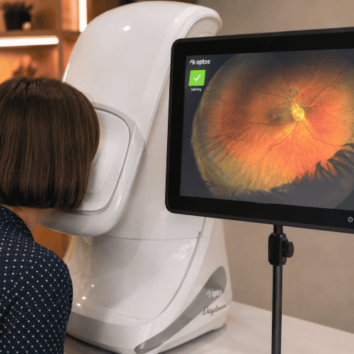
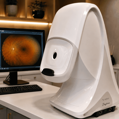
While OCT allows us to look through the layers of the macula, Optomap gives us a much wider view across the retina. This helps us document the appearance of the macula and the surrounding retina and provides an image that can be compared with future examinations.
Optomap also allows us to use autofluorescence imaging, which can reveal changes in the health and activity of the retinal pigment epithelium (RPE) – the layer of cells that supports the light-sensitive retina. In AMD, changes in autofluorescence can help highlight areas where these cells are becoming stressed or damaged, sometimes extending beyond what is immediately obvious on a standard retinal image.
Used alongside OCT, it gives us another way of looking at, recording and monitoring changes associated with AMD over time.
WHAT HAPPENS NEXT?
What happens if we find AMD
Finding signs of AMD does not necessarily mean that you need treatment. What happens next depends on the type of AMD, how advanced the changes are and whether they appear stable or are progressing.
If we find early or stable changes, we may recommend monitoring your eyes with regular examinations and imaging so that we can compare them over time. We will explain what we have found, discuss any changes you should look out for and advise you on ways to support your eye health.
If we see signs that suggest wet AMD or another change requiring specialist assessment, we will arrange an appropriate referral. Wet AMD can sometimes progress quickly, so prompt assessment is important as treatments are available that can help protect vision.
WORRIED ABOUT YOUR VISION
Has your vision suddenly changed?
A sudden change in your central vision needs prompt attention. This might include straight lines suddenly appearing wavy or distorted, a new blurred or dark area in the centre of your vision, or a noticeable difference between your two eyes.
These symptoms can sometimes be associated with wet AMD, where early assessment is important because treatment may be needed quickly.
If you notice a sudden change, don’t wait for your next routine eye examination – seek urgent advice.
How is wet AMD treated?
Wet AMD can often be treated with medicines called anti-VEGF drugs, which are given by injection into the eye by a specialist ophthalmology team. These medicines work by reducing the activity of abnormal blood vessels beneath the retina and helping to control leakage and swelling.
The thought of an injection into the eye can understandably sound daunting, but anaesthetic drops are used to numb the eye beforehand, and most people experience little or no pain during the procedure.
Treatment has transformed the outlook for people with wet AMD. Although it cannot restore every change that has already occurred, it can often stabilise vision and, in some people, improve it. Treatment usually involves a series of injections followed by ongoing monitoring, with the frequency tailored to how the eye responds.
This is why recognising a new change in vision and arranging prompt specialist assessment is so important.

What can I do to reduce my risk of AMD?
You cannot completely prevent AMD. Age is the biggest risk factor, and our genes also play a part. Having a close relative with AMD can increase your risk, but AMD is not usually inherited in a simple way, having a parent with AMD does not mean that you will inevitably develop it too. Many different genes appear to influence susceptibility, alongside factors such as smoking, diet and general health.
We cannot change our age or the genes we inherit, but there are things we can do to reduce the risks we can control and help protect the health of the macula.
Don’t smoke

Don’t Smoke
Eat well

Eat well
Look after your general health

Live Well


Understanding your eyes matters
If you are concerned about AMD, have a family history of the condition, or simply want to understand more about the health of your eyes, we are here to help.
Our enhanced eye examinations give us additional time to discuss your individual risk factors and examine your eye health carefully. Advanced imaging such as OCT and Optomap can provide further information where appropriate, while allowing us to build a detailed record of your eyes that can be compared over time.
Most importantly, we will explain what we find, what it means for you and whether anything further needs to happen.