
Ultra Widefield Retinal Imaging (Optos)
What Is an Optomap Retinal Scan?
A much wider view of your retina
The retina lines the inside of your eye and plays a vital role in your vision. Examining it carefully is an important part of understanding the health of your eyes.
The difficulty is that the retina extends much further around the inside of the eye than we can see through the pupil in a single conventional view.
Optomap ultra-widefield imaging allows us to see much more.
In just a few moments, it captures a remarkably wide image of the retina, including areas towards the periphery that can otherwise be difficult to examine and document.
Optos widefield imaging is part of our enhanced eye examination at Jacksons Styling Opticians in Nantwich.
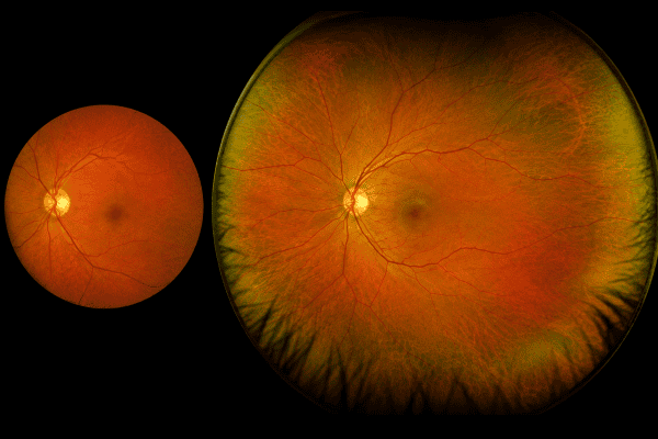
A standard fundus photograph compared with a digital widefield image
See the retinal periphery
A standard retinal photograph gives us a useful view of the central part of the retina, but it only captures a relatively small area at any one time. It’s a little like looking into a room through a keyhole – you can see what’s directly in front of you, but not what’s happening around the edges. Optomap is more like opening the door.
Its ultra-widefield technology allows us to see much further across the retina and out towards the periphery, giving us a far broader view of your eye health in a single image.
Why do we want to see the peripheral retina?
Not every important change happens near the centre of your vision.
Some retinal conditions can begin or show signs towards the outer edges of the retina, where you may be completely unaware that anything is happening.
Optomap gives your optometrist a widefield view that can help us identify and document features such as retinal holes or tears, areas of retinal degeneration, changes to blood vessels, pigmentation, naevi and other abnormalities within the peripheral retina.
If we see something that requires a closer look, we can then investigate it further or arrange referral when appropriate.
More than simply taking a photograph
Capturing the image is only the beginning.
Your optometrist examines your Optomap image as part of your overall eye examination, considering what we see alongside your symptoms, vision, prescription, medical and family history, and everything else we have discovered about your eyes.
We can also show you the image.
For many patients, seeing their own retina, including their optic nerve, macula and blood vessels, makes it much easier to understand what we are looking at and why it matters.
A valuable record of your eyes
One of the biggest advantages of digital retinal imaging is being able to keep the images and compare them in the future.
Your first Optomap provides us with a detailed baseline of what your retina looks like today.
When you return, we can compare your new images with previous ones. A feature that has remained unchanged for years may be reassuring, while a subtle difference from an earlier image may tell us that something deserves further investigation.
Sometimes it is the change over time, rather than how something looks on a single day, that provides the most useful information.
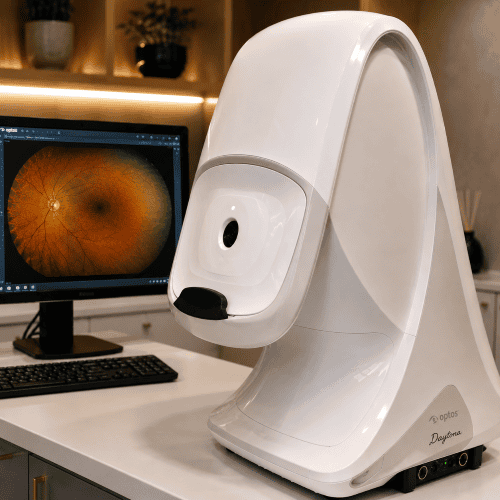
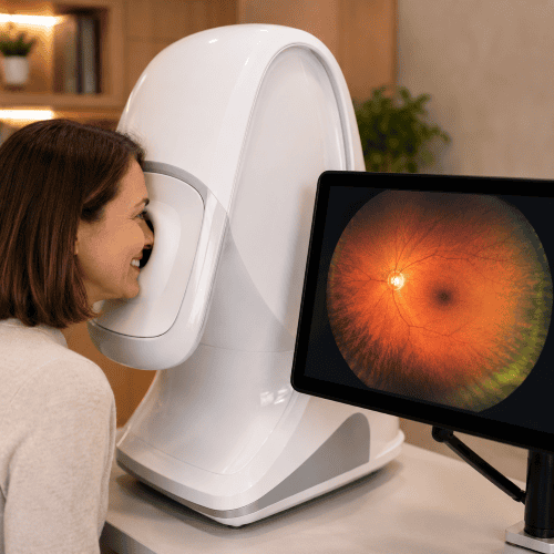
The Optos Daytona
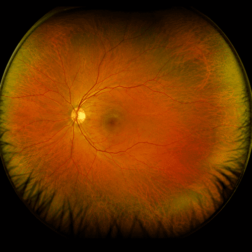
A widefield retinal image
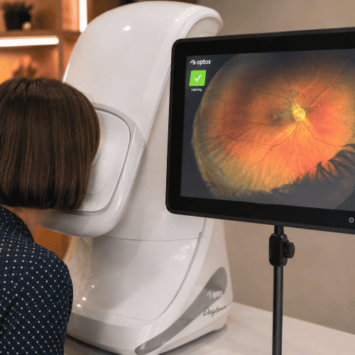
A patient undergoing Optos imaging
What can an Optomap image help us see?
Because Optomap captures such a wide area of the retina, it can help us examine and document many different features.
These may include
- retinal holes, tears and areas of degeneration
- changes in the retinal blood vessels
- haemorrhages or other signs of retinal disease
- retinal pigmentation
- choroidal naevi, sometimes described as “freckles” at the back of the eye
- changes associated with diabetes and other systemic conditions
- the appearance of the optic nerve and macula
- unusual features that we may want to monitor over time
An Optomap image does not diagnose these conditions by itself. It gives your optometrist another valuable source of information that can be interpreted alongside the rest of your eye examination.
What happens during an Optomap scan?
Having an Optomap image taken is quick, painless and non invasive.
You simply look into the instrument while it captures an ultra widefield image of your retina. Nothing touches your eye and the image itself is captured extremely quickly.
For many people, we can obtain this wide view without dilating the pupils.
There will still be occasions when your optometrist recommends dilating drops so that we can examine your retina directly in more detail. Optomap doesn’t replace clinical examination when that is needed.
Is Optomap the same as OCT?
No. And this is an important difference.
Optomap shows us across the retina. OCT shows us through it.
Optomap gives us an exceptionally wide view of the retinal surface, including the peripheral retina.
OCT gives us highly detailed cross sectional images through the retinal layers and allows us to take precise measurements of structures such as the macula and retinal nerve fibre layer.
Neither makes the other redundant.
Used together, they give us two very different perspectives on the health of your eyes.
Why we use Optomap at Jacksons
We don’t use technology simply because we have it. We use it when it gives us useful information about your eyes.
Optomap allows us to examine and document a much wider area of your retina, OCT allows us to investigate its microscopic structure, and your optometrist brings all of that information together with the findings from the rest of your examination.
It means we’re not relying on a snapshot in time either. We are building a detailed record of your eyes that we can return to and compare year after year.
A wider view. A better understanding of your eyes.
Our advanced eye examinations give us the time and technology to look carefully at your eyes, explain what we find and keep a detailed record that can be compared at future visits.
Book your eye examination at Jacksons Styling Opticians in Nantwich.