
Common Eye Disease
Glaucoma
Understanding a condition that can gradually damage the optic nerve
Glaucoma?
Glaucoma is the name given to a group of eye conditions that cause progressive damage to the optic nerve – the nerve that carries visual information from your eye to your brain.
The damage usually happens slowly and can initially affect your peripheral, or side, vision. Because the central vision often remains clear until much later, and one eye can compensate for the other, glaucoma can develop for a long time without you being aware that anything has changed.
Eye pressure is an important part of glaucoma, but the condition is not simply caused by having high pressure in the eye. Some people develop glaucoma despite having pressures within the normal range, while others have higher than average pressures without developing optic nerve damage.
This is why we assess several things together, including eye pressure, the appearance of the optic nerve, OCT imaging and your visual fields and why looking for changes over time can be particularly important.
Glaucoma cannot usually be cured, and vision already lost cannot be restored. However, when it is identified and treated appropriately, further damage can often be slowed or prevented, helping to preserve useful vision for the future.

What you might notice
One of the difficulties with glaucoma is that you may not notice anything at all in its early stages. There is usually no pain, and your central vision can remain clear while changes are developing elsewhere in your field of vision.
Glaucoma often begins by affecting peripheral, or side, vision. These changes tend to happen gradually, and because your brain is remarkably good at filling in missing information and one eye can compensate for the other – significant damage can occur before you become aware of a problem.
As glaucoma becomes more advanced, you may begin to miss objects to the side, find navigating unfamiliar places more difficult or notice areas of your vision that seem less clear. Eventually, central vision can also be affected.
This is why glaucoma is so often detected during a routine eye examination rather than because someone has noticed a change in their sight. Regular examinations are particularly important if you have other risk factors for the condition.
What is happening inside the eye?
At the back of your eye is the optic nerve, made up of more than a million tiny nerve fibres that carry visual information from the retina to your brain. These nerve fibres are essential for sight.
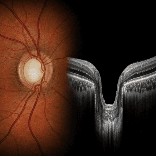
In glaucoma, some of these fibres gradually become damaged and are lost. As this happens, the appearance of the optic nerve can change. The central area of the nerve, known as the optic cup, may become larger or deeper, while the surrounding healthy nerve tissue becomes thinner.
The layer of nerve fibres across the retina also becomes thinner. This is particularly useful for us to examine with OCT imaging, because OCT can measure these structures in remarkable detail and help us look for subtle changes over time.
As more nerve fibres are lost, corresponding areas of the visual field can also be affected. This is why examining the optic nerve, OCT imaging and visual field testing provide different pieces of the same picture when we are looking for glaucoma.
SEEING BENEATH THE SURFACE
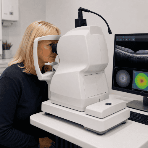
OCT allows us to examine and measure the delicate structures around the optic nerve, including the retinal nerve fibre layer. In glaucoma, characteristic areas of this layer can become thinner as nerve fibres are gradually lost.
A single OCT scan gives us useful information, but one of its greatest strengths is what happens when we repeat the scan over time. By accurately comparing today’s measurements with previous examinations, we can look for subtle patterns of thinning and establish whether something is genuinely changing or has simply always been normal for your eyes.
OCT can sometimes identify structural change before a corresponding change is detected on a visual field test, making it particularly valuable when monitoring people who may be at increased risk of glaucoma.
However, an OCT scan does not diagnose glaucoma by itself. We interpret the findings alongside the appearance of your optic nerves, eye pressures, visual fields, family history and other individual risk factors to build a much more complete picture.
Eye pressure matters – but it is only part of the picture
The pressure inside your eye is known as intraocular pressure (IOP). Maintaining some pressure is necessary to keep the eye healthy and maintain its shape, but higher eye pressure is an important risk factor for developing glaucoma.
There isn’t, however, a single pressure at which glaucoma suddenly develops. Some people have higher than average eye pressures and never develop optic nerve damage, while others develop glaucoma even though their pressures are within what would traditionally be considered the normal range.
Eye pressure can also vary naturally during the day, which is another reason why we don’t interpret a single measurement in isolation.
Instead, we consider your eye pressure alongside the appearance of your optic nerves, OCT measurements, visual fields, corneal thickness, family history and other individual risk factors. It is the combination of these findings, and particularly how they change over time – that helps us understand your glaucoma risk.


Understanding your visual field
WHAT HAPPENS NEXT?
What happens if we find something suspicious?
Finding an unusual optic nerve appearance, raised eye pressure or an OCT measurement outside the expected range does not necessarily mean that you have glaucoma. There is considerable natural variation between people’s eyes, which is why we consider all of the findings together.
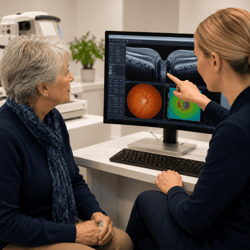
Sometimes the most useful next step is to repeat measurements or monitor your eyes over time. This allows us to establish what is normal for you and look for genuine change rather than making a judgement from a single examination.
If the combination of findings suggests that glaucoma may be present, or that your risk is sufficiently increased, we will explain what we have found and arrange an appropriate referral for further assessment.
The important thing is that glaucoma is often identified before you are aware of any problem with your vision. Detecting and monitoring suspicious changes gives us the opportunity to make sure treatment is started when it is needed.

WHAT HAPPENS NEXT?
How is glaucoma treated?
Who is at greater risk of glaucoma?

Can I reduce my risk of sight loss from glaucoma?
There is no guaranteed way to prevent glaucoma, and some of the most important risk factors – such as age, genetics and family history, are things we cannot change. What we can do is improve the chances of detecting glaucoma early and limiting the damage it causes.
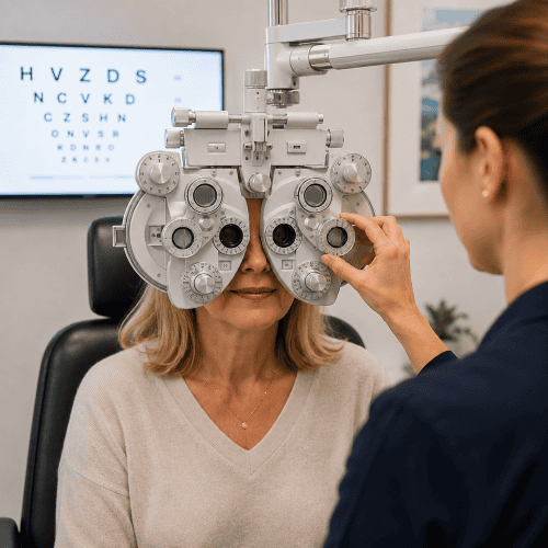
Have regular eye examinations
Glaucoma can develop without causing noticeable symptoms, so regular examination of your eye pressures and optic nerves is important, particularly if you have additional risk factors.
Know your family history
If a parent, sibling or other close relative has glaucoma, tell your optometrist. It may influence how we interpret your findings and how closely your eyes should be monitored.
Attend your monitoring appointments
If you have been diagnosed with glaucoma or ocular hypertension, regular follow-up is important even when your vision seems completely normal. Comparing eye pressures, OCT scans and visual fields over time helps establish whether your condition remains stable.
Use treatment as prescribed
If you have been prescribed glaucoma eye drops, using them regularly is important. Glaucoma treatment is designed to protect your vision for the future, so you may not notice any immediate difference when you use the drops.
Look after your general health
Keeping active, eating a balanced diet and managing conditions such as high blood pressure and diabetes are important for your overall health as well as your eyes. However, a healthy lifestyle is not a substitute for glaucoma treatment or monitoring if these are needed.
The reassuring message is that most people whose glaucoma is identified and treated appropriately retain useful vision throughout their lives. Early detection and continued monitoring give us the best opportunity to protect your sight.
Glaucoma is often found before you notice it
ecause glaucoma can develop gradually without causing obvious symptoms, regular eye examinations play an important part in identifying people who may be at increased risk.
Our enhanced eye examinations allow us time to look carefully at your optic nerves, measure your eye pressures and consider your individual risk factors. Where appropriate, OCT imaging and visual field testing provide additional information and, importantly, allow us to compare findings over time.
No single test tells us everything about glaucoma. By bringing the different pieces of information together, we can build a much clearer picture of what is normal for your eyes and whether anything appears to be changing.
If we find something that needs further investigation, we will explain what we have found and what needs to happen next.

Understanding your eyes matters
If you are concerned about glaucoma, have a family history of the condition, or simply want to understand more about the health of your eyes, we are here to help.
Our enhanced eye examinations give us additional time to discuss your individual risk factors and examine your eye health carefully. Advanced imaging such as OCT and Optomap can provide further information where appropriate, while allowing us to build a detailed record of your eyes that can be compared over time.
Most importantly, we will explain what we find, what it means for you and whether anything further needs to happen.
Further trusted information
For more detailed information about glaucoma, including diagnosis, monitoring and treatment, we recommend information from the NHS and Glaucoma UK.
Clinically reviewed by Helen Corson, BSc MCOptom PGCert IP
Last reviewed: August 2026