Optical Coherence Tomography – OCT
What is OCT?
Optical Coherence Tomography – OCT, is a specialist scan that, until recently, was only found in specific hospitals across the UK. An OCT creates a 3D image of the back of your eye (the retina) and allows our trained optometrist to look at multiple layers of your retina in microscopic detail. To create the 3D image an OCT scan uses a laser to scan your retina, therefore nothing touches or blows on your eye, this means that we can use an OCT scan on everybody, no matter how old or young you are.
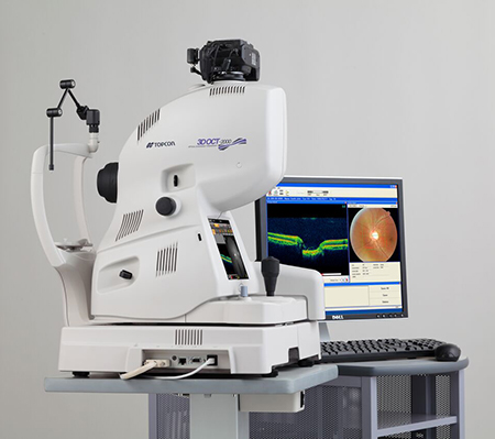
How does an Optical Coherence Tomography Work
An OCT uses a scanning laser to examine the retina. It inputs the results of these scans into a piece of computer software and the scans are converted into a fully interactive 3D image. The scans can be compared to a cake. When we look into the eye, we normally see only the surface of the cake – the retinal surface. With OCT we can take a slice out of the cake to see all the different layers that make up the retina, focus on a certain sections and pinpoint any unhealthy changes or abnormalities.
What’s the difference between retinal photography and Optical Coherence Tomography scanning?
Fundus photography takes a photograph of the surface of your retina only. The OCT scan takes a 3D scan of your entire retinal thickness allowing our optometrist to “peel back” each layer until they find the cause of the abnormality and can quickly diagnose any problems or areas for concern.
Should I have an Optical Coherence Tomography?
We recommend that every patient has an initial baseline OCT scan. This will be stored by our software and compared to any future OCT scans for minor, but significant changes to your retina. An OCT scan is particularly useful for any patients who have concerns of Glaucoma, Macular Degeneration and “Flashes and Floaters”.
OCT scanning is included within our Enhanced Eye Health Examination and is available as an upgrade for patients qualifying for and NHS funded Sight Test.
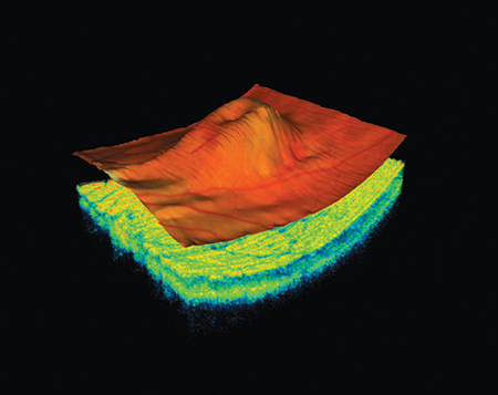
What do the scans look like?
OCT scan of Wet AMD. This scan shows fluid from leaking blood vessels trapped underneath the surface of the retina. The blood would not be visible on examination by more conventional techniques. The patient may have some reduction in their vision but symptoms at this stage can be vague. Recent advances in treatment mean that the vision in cases like this can be saved using intraocular injections.
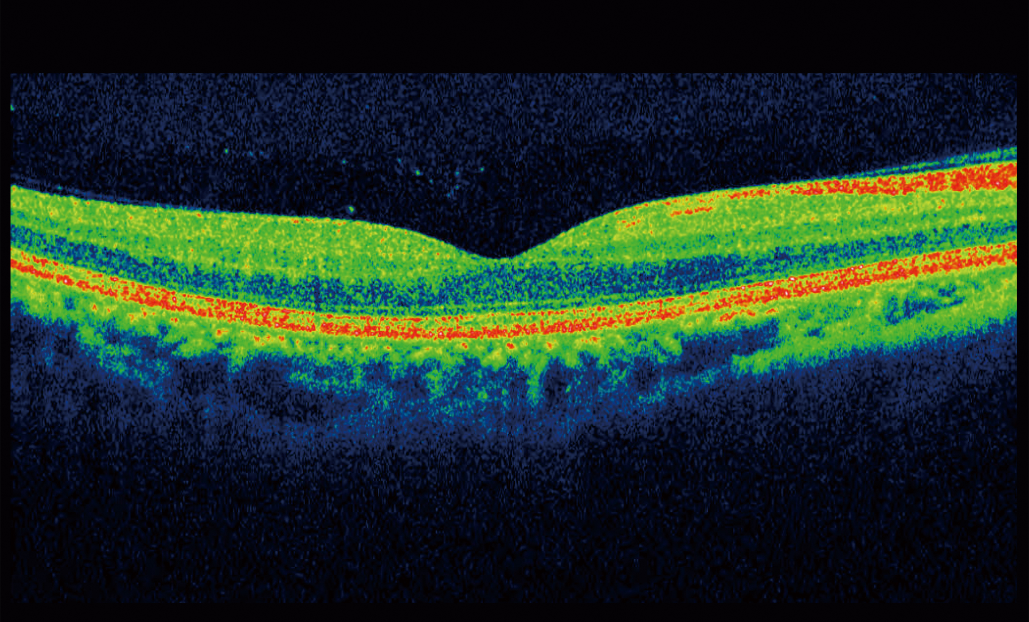
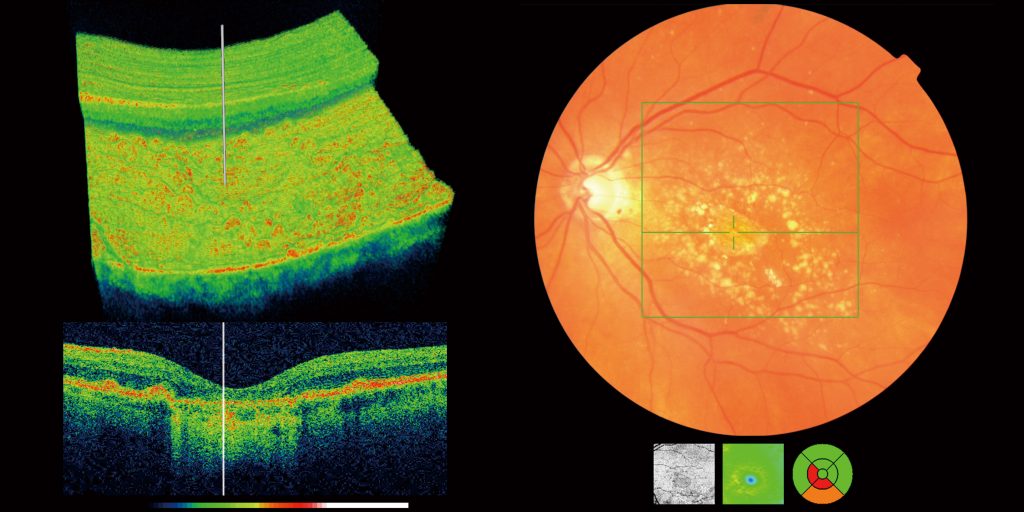
OCT scan of a healthy macula. Contrast this with the scan above. All the retinal layers are regular and there is no intraretinal fluid or cystic spaces.
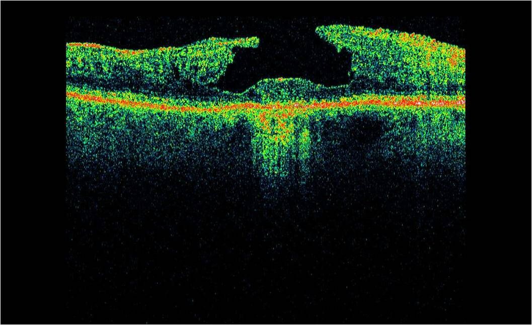
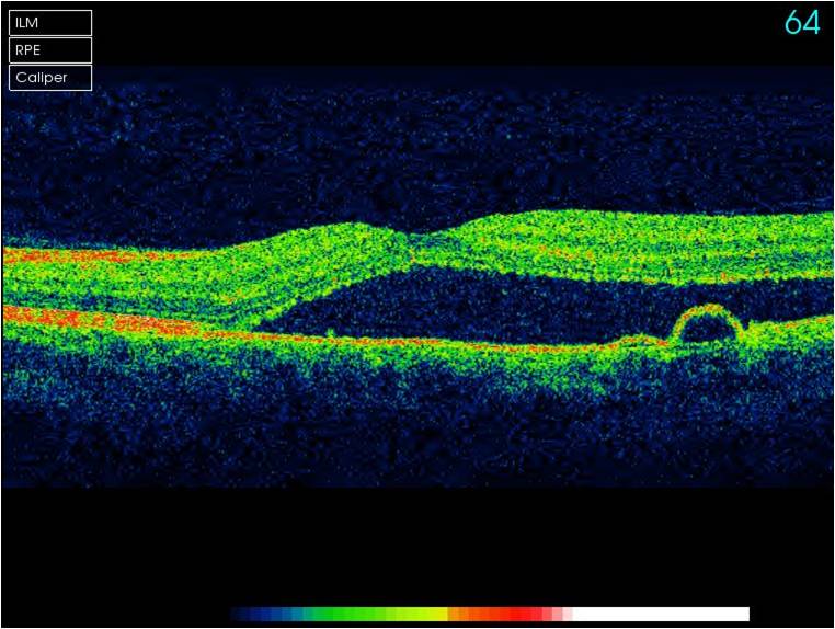
OCT scan of a macula hole. OCT makes the diagnosis of macula hole much easier than by direct viewing alone. With modern technological advances macula holes are treatable if caught in time.
Sample Videos
To see what our OCT scans look like, please watch the videos below.