
Why We Always Recommend OCT and Optomap as Part of Every Eye Examination
When you visit Jacksons Styling Opticians – Nantwich, you’re not just having a sight test — you’re having a comprehensive assessment of your eye health.
Our eyes are extraordinary structures, and many sight-threatening diseases begin without any pain or early warning signs. That’s why we recommend including OCT (Optical Coherence Tomography) and Optomap ultra-widefield imaging as part of every eye examination.
These technologies allow us to look deeper, wider, and earlier than ever before — giving you the highest standard of preventative eye care available.
OCT: Seeing beneath the surface
OCT scanning uses light waves to create a 3D, cross-sectional image of the retina and optic nerve — revealing microscopic details invisible to standard examination.
We can identify and monitor:
-
Macular degeneration (AMD) – early drusen deposits or fluid changes, before vision is affected
-
Epiretinal membranes – scar tissue that distorts the retina
-
Macular holes and vitreomacular traction – which can cause sudden central vision loss
-
Diabetic macular oedema (DMO) – fluid leakage that can occur even when diabetic control seems stable
-
Glaucoma – thinning of the retinal nerve fibre layer years before field loss occurs
-
Optic neuritis, papilloedema, or vascular occlusions – conditions linked to inflammation, raised pressure, or circulation problems
Because OCT provides precise, repeatable 3D images, we can compare scans year to year and spot even the smallest change.
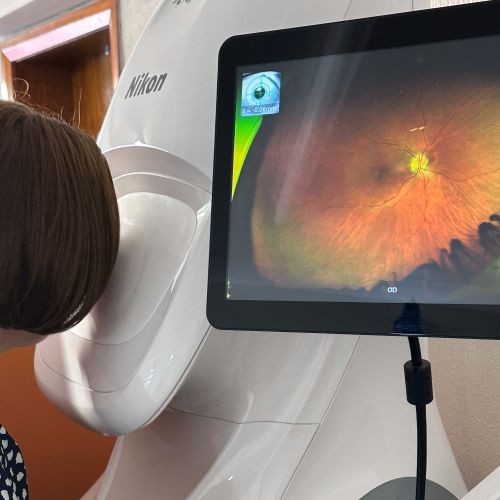
Optomap: The panoramic view of your retina
While OCT lets us look through the retina in exquisite detail, Optomap shows us an ultra-widefield image of up to 200° — more than four times the area seen with a standard fundus camera.
It helps detect:
-
Peripheral retinal holes or tears before they lead to detachment
-
Lattice degeneration and other structural weaknesses
-
Diabetic or hypertensive retinopathy affecting the peripheral retina
-
Choroidal naevi (pigmented lesions) that require ongoing monitoring
-
Signs of inflammation or vascular occlusion linked to systemic conditions
This panoramic view often reveals changes that would otherwise go unnoticed until they caused visual symptoms.
Why this isn’t covered by the NHS sight test
Many patients are surprised to learn that OCT and Optomap scans are not included in an NHS eye examination.
This is because the NHS sight test is designed primarily to:
-
Check your visual acuity and prescription
-
Assess the external and internal appearance of the eyes using standard equipment
-
Detect obvious signs of eye disease
While this provides an important baseline, the NHS test does not include advanced imaging such as OCT or ultra-widefield retinal scanning, as these are not currently funded under the NHS General Ophthalmic Services (GOS) regulations.
In contrast, private enhanced eye examinations like ours go far beyond the basic sight test.
By incorporating OCT and Optomap imaging, we can detect disease at the earliest, most treatable stages, often before symptoms develop — helping you avoid irreversible damage and providing peace of mind that your eyes have been examined in full detail.
It’s the difference between a snapshot and a high-definition scan — both valuable, but one gives you far more information.
An investment in your long-term eye health
Both OCT and Optomap scans are:
-
Quick and completely painless
-
Safe for all ages
-
Performed in just a few minutes
We review the results with you immediately, explaining what we see and what it means for your individual eye health.
Including these scans as part of your routine care is one of the most effective ways to protect your vision for life.
Book your advanced eye examination
If it’s been over a year since your last appointment — or you’ve never experienced OCT or Optomap imaging before — we’d strongly recommend booking an enhanced eye examination.
Telephone us on 01270 625889
Book an appointment at Jacksonsopticians.com/book
Visit us at Jacksons Styling Opticians – 43–45 Welsh Row, Nantwich CW5 5EW